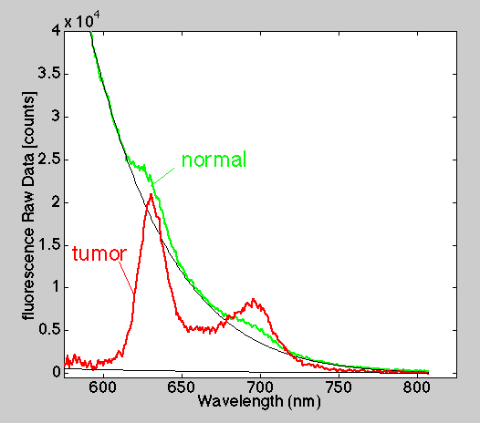
Figure 1: Typical raw fluorescence measurements.
|
PDT NewsEtc. | Raw fluorescence spectra |
Table of Contents | PREVIOUS PAGE | NEXT PAGE
Figure 1 shows typical raw fluorescence measurements made with a single bare optical fiber which delivers excitation and collection fluorescence emission. The green lines are 3 normal esophageal tissue sites. The red lines are 3 esophageal tumor sites. The black lines are the tails of Gaussian curve fits which mimic the background autofluorescence of the tissue.
Figure 2 shows the photosensitizer (Photofrin) spectra after subtracting the Gaussian curves that characterizes the background autofluorescence.
Figure 1: Typical raw fluorescence measurements.
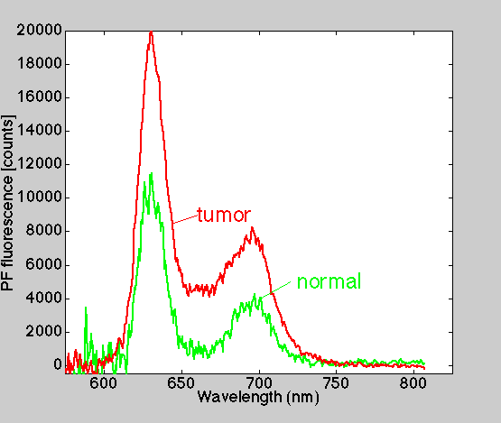
Figure 2: Fluorescence of photosensitizer (Photofrin) after subtraction of background autofluorescence.
Table of Contents | PREVIOUS PAGE | NEXT PAGE