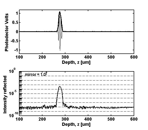

Calibration was achieved by comparison with the intensity signal from a glass/oil interface (reflectivity of intensity = 4x10⁻⁴), using the strong signal at the focus of the objective lens.

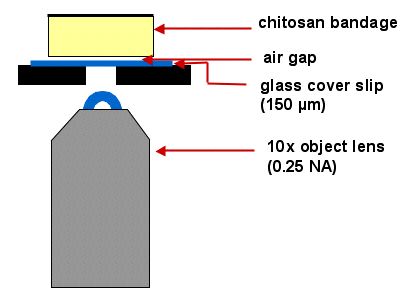
The light passes through an objective lens and glass coverslip to reach the bandage from below.

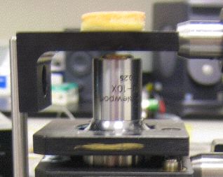
Photograph of the objective lens and bandage.

Focus tracking: We did not implement true focus tracking, but stitched together several images in which the focus of the objective lens, where OCT signal strength is maximal, had been translanted to a new depth in the bandage. The system generated 10 x-z images of the chitosan bandages. For each image, the focus of the objective lens (10x, NA = 0.25) was translated by 1/10th of the full axial range of imaging, so as to move the focus of the objective lens to 10 uniformly separated positions. Then the best portions from each of the 10 images were stitched together to yield a single image which was minimized the spatial variations in the strength of the focus of the objective lens.
Bandage preparation: Two types of bandages were tested. One type was a "good" bandage whose preparation involved a freeze-drying protocol that yielded vertical lamina. The second type was a "bad" bandage whose preparation yielded horizontal lamina.