Slide 7/23

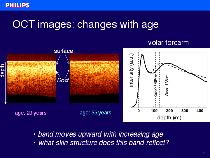
The cross-sectional OCT images in this slide are measured on the forearm of a 23 year-old volunteer (left image) of a 57 year-old volunteer (right image). In the OCT images two bright bands can be observed, they are roughly indicated in the images by the arrows. The first bright band is due to the reflectance from the stratum corneum. Just below this layer, in the viable epidermis, the signal intensity diminishes. As will be discussed later on, the second reflecting layer, deeper in the skin, can be ascribed to back scattering of light at fibrous structure of collagen in the dermis. In the younger volunteer this reflecting layer is located much deeper below the surface as compared to the situation in the elderly volunteer.For analysis, the surface in the image was flattened, (as can be seen in the slide). An average intensity profile was calculated from the integration of the signal along the lateral position x as a function of depth. An intensity profile shows two peaks, caused by the two bright reflecting bands. The first peak, due to the skin surface, was set to a depth of 0 mm. The location of the second peak at Doct is determined relative the skin surface. The profiles and images show that the value of Doct is relatively small for the older volunteer. Dashed line profile: older volunteer, solid line profile: younger volunteer.
index | previous | next

