Slide 22/23
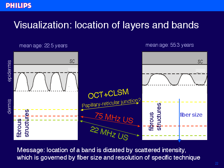
This slides gives a visual representation of the dermal structures measured by the different techniques. Again, with the optical techniques the dermal band moves up with age. The transition is most probably due to the papillary-reticular junction. On the other hand with ultrasound the band moves down with age. Apparently, the location of a band is dictated by scattering intensity, which is governed by fiber size and resolution of the specific technique.
index | previous | next