Slide 15/23

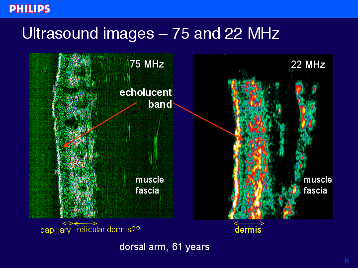
In this slide two ultrasound images are shown, measured in a 61 years old volunteer. The left image is measured with 75 MHz ultrasound, the right one with 22 MHz. In both images, the surface of the skin is seen as a bright reflecting layer, below this entry echo a darker, echo-lucent structure is visible, followed by an again echo-rich region. The transition between the dermis and subcutaneous fat layer is nicely visible in both images. The following question arises: Are the same transitions seen with both ultrasound frequencies?
index | previous | next

