© 1998 Steven L. Jacques, Scott A. Prahl
Oregon Graduate Institute
 |
ECE532 Biomedical Optics © 1998 Steven L. Jacques, Scott A. Prahl Oregon Graduate Institute |
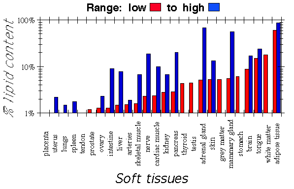
Soft tissue optics are dominated by the lipid content of the tissues. Such lipid constitutes the cellular membranes, membrane folds, and membranous structures such as the mitochondria (about 0.5 µm). While other objects such as protein aggregates and the nucleus are also sources of scattering, the lipid/water interface of membranes presents a strong refractive index mismatch and so plays a major role is scattering. The following graph summarizes the lipid contents of various tissues:

Mie theory provides a simple first approximation to the scattering of soft tissues [ref. 2]. The approximation involves a few assumptions:
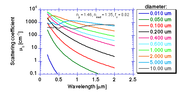
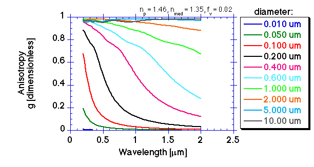
In the following graphs, a 2% volume fraction of lipid is packaged as spheres of one size, for a series of choices of sphere diameter (radius a), adjusting the number density to maintain a constant volume fraction:


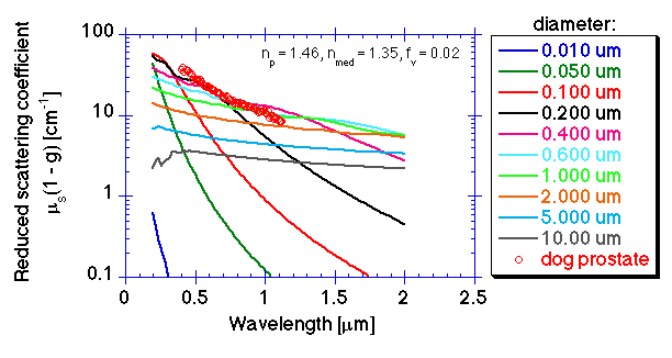
This picture includes the experimental values of µs(1 - g) for dog prostate, illustrating that the prostate is mimicked by a 2% volume fraction of lipid spheres in the 0.200-0.600 µm diameter range. Other soft tissues vary only a little from this general pattern for prostate tissue.

The wavelength dependence of light scattering in soft tissues such as the prostate suggests that cellular structures equivalent to spheres with diameters in the range of 0.200-0.600 µm contribute most of the scattering. The magnitude of the scattering suggests that a volume fraction of 0.02 or 2%, which is roughly the reported lipid content of prostate and many other soft tissues, yields a magnitude for µs(1 - g) which matches the magnitude of µs(1 - g) for prostate. Deviations from our assumed values for np based on lipid and nmed based on cytosol and from our assumed value for fv will affect the magnitude of µs(1 - g). But in general,soft tissues with higher (or lower) lipid content will show increased (or decreased) scattering, while the wavelength dependence of µs(1 - g) should not change greatly.