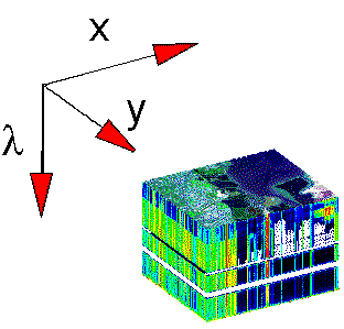
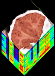
Hyperspectral imaging is the technique of acquiring two-dimensional images in which each pixel has spectral information. Such an image is sometimes called a data cube or hypercube, in which the x-y plane shows the tissue structure and the z-axis encodes the wavelength-dependent information.
One of the early examples in medical optics of hyperspectral imaging was the multifiber fluorescence probe developed by Michael Feld's group at M.I.T. to map atherosclerotic plaque. Each fiber in a multiple-fiber catheter sequentially delivered excitation light and collected fluorescent emission to yield a fluorescence spectrum for that pixel. The catheter had 19 fibers and hence the spatial resolution was coarse, however, the device could map the borders of artherosclertic plaque within arteries. Rebecca Richards-Kortum, Univ. of Texas-Austin, has been implementing fluorescence based hyperstatial imaging of the cervix to detect cervical dysplasia. Today video cameras are used rather than optical fibers to achieve high spatial resolution.
NASA and its various subcontractors have developed hyperspectral imaging based on reflectance spectra for satellite mapping of the planetary surface for weather, agriculture, and other tasks. Now this technology is being applied to medicine. This month's gallery shows some of the current work from Greg Bearman at JPL and collaborators at UCLA on hyperspectral imaging of brain tissue slices.
Further information on hyperspectral imaging can be found at these sites:
- Opto-Knowledge Systems, Inc. (OKSI): Nahum Gat and Suresh Subramanian,
- NASA: Greg Bearman, Jet Propulsion Laboratory (JPL), et al.
 Hyperspectral imaging is a 2-D image where each pixel has a full spectrum of information. "lambda" means signal at a particular wavelength. Nahum Gat and Suresh Subramanian, Opto-Knowledge Systems, Inc. (OKSI) link | Hyperspectral imaging.Each pixel of a 2-D image has a full spectrum of information, shown here as the z-axis labeled "lambda". |
 Click here to expand figure. Click here to expand figure.Hypercube of brain tissue slice. X and Y are the 2D spatial map and Z is the wavelength dimension. Image is located at Nahum Gat and Suresh Subramanian, Opto-Knowledge Systems, Inc. (OKSI) link. | Hypercube of brain tissue slice.JPL and UCLA are utilizing hyperspectral techniques to generate functional mapping of the brain. The idea is to identify areas of the brain that perform specific functions without doing histopathaology on every slice.Image was prepared by Gregory Bearman, JPL. See Greg's liquid crystal tunable filter spectrometer. |
NewsEtc. Index
OMLC home page