A simple optical fiber spectrometer was implemented one afternoon as a demonstration for some students. Hence, this data is rather noisy and certainly not optimized. Nevertheless, the data is sufficient to specify the optical properties of the skin.
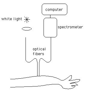
White light was focused into an optical fiber (600-μm-dia.), called the "source", that brought the light to the skin surface as the fiber contacted the skin. A second optical fiber (600μm diameter), called the "collector", was placed 2 mm to the side of the source fiber. The collector fiber collected light that entered the skin and diffused laterally in the skin, and brought that collected light to a spectral detector (an inexpensive commercially available unit) that used a diffraction grating to spread the light over a diode array detector, yielding a measurement of the spectrum from 400-900 nm. The spectrometer was connected to a computer that acquired the spectra.
Figure: Optical fiber spectrometer. Source and collector fibers separated by 2 mm.
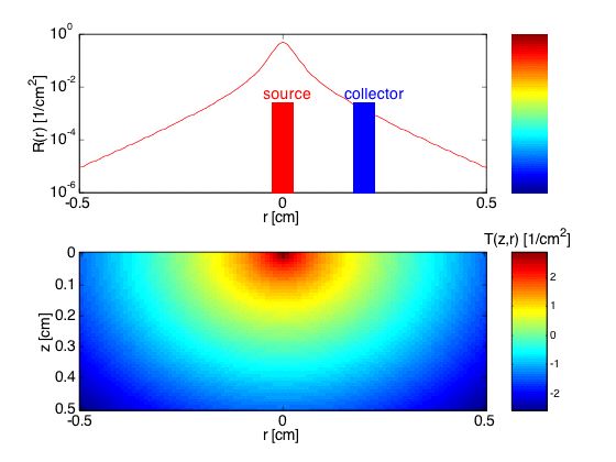
Figure: Collection of escaping light, R(r), at r = 0.2 cm. The transport factor T(z,r) [cm-2] is shown within the tissue (577 nm wavelength, blood volume = 0.5%, oxygen saturation = 70%, scattering = standard dermal scattering ---> μa = 1.38 cm-1, μs' = 32.5 cm-1).