A very practical question is "how dark is the pigmented epidermis of a skin site?" During pulsed laser treatments of portwine stain lesions and other vascular lesions, the heat deposition in the epidermis is a limiting factor. Epidermal pigmentation can be pertinent to laser removal of tattoos and laser hair removal as well.
Kollias and Baqer (1986) proposed that one could document epidermal pigmentation by the spectral behavior of skin reflectance in the 620-720 nm range. A measurement (Mskin) of light backscattered from the skin and a measurement (Mstd) of light backscattered from a reflectance standard of value Rstd are made. These measurements depend on the light source spectrum (S), the detector spectral response (D), the fraction of reflected light escaping a medium that is collected by the measurement system (f), and the true reflectance of the skin:
Mskin/Mstd = (SfskinRskinD)/(SfstdRstdD)
= Rskin Rstdfskin/fstd = Rskinf*
where f* = Rstdfskin/fstd, a calibration factor. Kollias and Baqer made measurements on a pigmented skin site and on a nearby vitiligo site which is devoid of melanin pigment. The ratio of these two measurements would be:
Mpigmented/Mvitiligo = Rpigmentedf*pigmented/(Rvitiligof*vitiligo) = Rpigmented/Rvitiligo
because f*pigmented = f*vitiligo and so cancels out. The f* is not significantly affected by a thin superficial absorbing layer (pigmented epidermis), fskin being dominated by the bulk tissue optical properties below the surface. So the ratio of true reflectances is obtained, and they differ only by the effect of the pigmented epidermis.
The optical density (OD) of the pigmented epidermis for photons passing twice through the epidermis, once upon entering the skin and once upon escape, is defined as:
OD = -log10(Rpigmented/Rvitiligo)
The following figure illustrates the OD attributed to the pigmented epidermis. Figure A compares a normal pigmented skin site and a vitiligo site. Figure B compares measurements of the more pigmented dorsal forearm and the less pigmented ventral forearm of 10 subjects, plotting -log10(Rdorsal/Rventral).
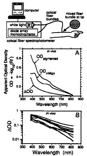
Figure: In vivo spectrometer measurements of pigmented epidermis.
(A) Comparison of normally pigmented and vitiligo skin site on arm.
(B) Comparison of dorsal and ventral forearm skin sites.
Figure from Jacques and McAuliffe (1991).
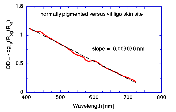
The following figure shows an OD spectrum based on data from Kollias and Baqer for a normally pigmented versus vitiligo site. The slope d(OD)/d(nm) is -0.003030 nm-1, where nm denotes wavelength in nanometers.
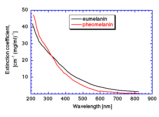
In the section of this article on the extinction coefficient, a graph of the extinction coefficient from Sarno and Swartz was presented in units of [cm-1 (mg/ml)-1]. For the in vivo spectrometer measurement, the photons have passed twice through the approximatley 60-µm thick epidermis, once entering and once escaping, so the pathlength is about 120 µm. Hence, the product of extinction coefficient and pathlength yields an optical density per unit concentration [(mg/ml)-1]. The extinction coefficient graph is redrawn below as an optical density graph per unit of eumelanin concentration.
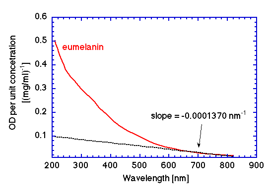
The slope between 620-720 nm is shown by a dashed black line. The value of the slope d(OD)/d(nm) is -0.0001370 [nm-1 (mg/ml)-1]. We can estimate the melanin concentration in the above in vivo pigmented site:
(-0.003030 nm-1)/(-0.0001370 [nm-1 (mg/ml)-1) = 21.9 mg/ml
Hence it is possible to assign a value of eumelanin concentration to a measured slope d(OD)/d(nm) of a pigmented epidermis. In this case, 21.9 mg/ml of eumelanin corresponded to a slope of -0.003030 nm-1.
Routine measurements with spectrometer
A routine measurement would be to simply measure a skin site Mskin, normalized by a measurement of Teflon MTeflon to cancel the light source (S) and detector (D) spectral dependencies. For convenience, let us call this normalized ratio X:
Mskin/MTeflon = fskinRskin / (fTeflonRTeflon) = X
In my experience with optical fiber spectrometers using Teflon as the reference standard, I have found that vitiligo sites, and extremely lightly pigmented Caucasians, present a 650-800 nm slope, d(X)/d(nm), that is relatively consistent from individual to individual. For our 2-mm-dia. mixed fiber bundle with Teflon as a reference standard, I found for vitiligo sites:
d(X)/d(nm) = -log10(Mskin/MTeflon) roughly equals -0.000500 nm-1
Therefore, one can routinely make measurements on individuals without a convenient vitiligo site and use this -0.000500 nm-1 value as a baseline for zero melanin. This baseline value will vary with the details of one's measurement device, but once determined it should be relatively consistent for all individuals unless there is significant edema or changes in skin thickness that affect the baseline reflectance spectrum.
In summary, the melanin content of the pigmented epidermis of skin can be conveniently characterized in units of mg/ml of eumelanin by measurements with an optical fiber spectrometer:
eumelanin [mg/ml] = ( d(X)/d(nm) - (-0.000500) )/(-0.0001370)
REFERENCES:
- N Kollias, A Baqer. On the assessment of melanin in human skin in vivo. Photochem. Photobiol. 43:49-54, 1986.
- SL Jacques, DJ McAuliffe. The melanosome: threshold temperature for explosive vaporization and internal absorption coefficient during pulsed laser irradiation. Photochem. Photobiol. 53:769-775, 1991.