Consider two approaches toward measuring the absorption coefficient of melanosomes:
- (1) measure the optical transmission through individual melanosomes using a microscope and calculate µa from the attentuated transmission.
- Goldman (1969) measured µa = 4100 cm-1 for retinal pigmented epithelium melanosomes at 694 nm.
- Sliney and Palmisano (1968) measured µa = 310 cm-1 for cutaneous melanosomes at 694 nm.
- Jacques and McAuliffe (1991) measured the difference in reflectance from ventral and dorsal sites of the forearm of 10 humans. The dorsal site was more pigmented due to sun exposure. The optical density associated with the difference in pigmentation between the sites was calculated: OD = -log10(Rventral/Rdorsal). The average difference optical density of the 10 humans was scaled by 3302 cm-1/OD to align the spectrum with the data generated using the explosive vaporization criterium.
- (2) measure the threshold pulsed laser radiant exposure that causes explosive vaporization of melanosomes and deduce µa.
- Jacques and McAuliffe (1991) used explosive vaporization to measure the µa of melanosomes in ex vivo human skin specimens, and the literature data for similar in vivo measurements. A Q-switched pulsed ruby laser (40 ns pulse duration) irradiated pigmented skin samples causing the melanosomes to explosively vaporize which caused a temporary whitening of the skin due to microcavity formation. In a few minutes, fluid refilled the cavities and the whitening disappeared. This phenomena had been studied by various investigators using a variety of pulsed lasers operating from UV to NIR. Jacques and McAuliffe demonstrated that the threshold temperature for this effect was about 112 degC. For example, a typical threshold radiant exposure for the effect at 694 nm in moist skin specimens resting at 20 degC was about Hth = 1.0 J/cm2. The relationship between Hth and the temperature jump dT is:
dT = (µaHth)/(rho C)
where rho is the density and C is the heat capacity of the melanosome (estimate: the product (rho C) = 3.22 [(J/cm3)/degC] for melanosomes with 54% water content). The dT was (112 - 20) = 92 degC. A typical µa was calculated:
µa = (rho C)(dT)/Hth = (3.22)(92)/(1.0) = 296 cm-1
Similarly, they used literature values for Hth for various pulsed lasers at various wavelengths to yield a spectrum of µa. - Jacques, Glickman, and Schwartz (1996) used explosive vaporization to measure the µa of melanosomes isolated from the bovine retinal pigmented epithelium. They used the Q-switched 532-nm doubled Nd:YAG laser and deduced µa = 2370 cm-1 for retinal melanosomes.
- Jacques and McAuliffe (1991) used explosive vaporization to measure the µa of melanosomes in ex vivo human skin specimens, and the literature data for similar in vivo measurements. A Q-switched pulsed ruby laser (40 ns pulse duration) irradiated pigmented skin samples causing the melanosomes to explosively vaporize which caused a temporary whitening of the skin due to microcavity formation. In a few minutes, fluid refilled the cavities and the whitening disappeared. This phenomena had been studied by various investigators using a variety of pulsed lasers operating from UV to NIR. Jacques and McAuliffe demonstrated that the threshold temperature for this effect was about 112 degC. For example, a typical threshold radiant exposure for the effect at 694 nm in moist skin specimens resting at 20 degC was about Hth = 1.0 J/cm2. The relationship between Hth and the temperature jump dT is:
The following figure summarizes the results from the above studies.
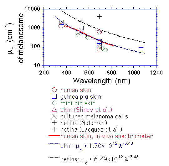
Figure: The absorption coefficient (µa [cm-1]) of the melanosome interior. Data based on threshold for explosive vaporization of melanosomes by various pulsed lasers (from Jacques and McAuliffe 1987 and Jacques et al. 1996), except for data of Goldman and data of Sliney et al. based on optical measurements. The solid red line is based on an in vivo optical fiber spectrometer measurement scaled to match data. The dashed blackline is the approximate expression mentioned at the bottom of this page.
A key lesson:
The concentration of melanin within melanosomes is quite variable. Ten-fold variation is to be expected. However, the general shape of the melanosome absorption spectrum is approximated:
µa = 1.70x10¹² nm-3.48 [cm-1] for skin
µa = 6.49x10¹² nm-3.48 [cm-1] for retina
where nm refers to the wavelength expressed in nanometers.
REFERENCES:
- L Goldman, The Skin, Arch. Environmental Health, 18:435, 1969.
- DH Sliney, WA Palmisano. The evaluation of laser hazards. AIHA Journ. 20:425, 1968.
- SL Jacques, DJ McAuliffe. The melanosome: threshold temperature for explosive vaporization and internal absorption coefficient during pulsed laser irradiation. Photochem. Photobiol. 53:769-775, 1991.
- SL Jacques, RD Glickman, JA Schwartz. Internal absorption coefficient and threshold for pulsed laser disruption of melanosomes isolated from retinal pigment epithelium. SPIE Proceedings 2681:468-477, 1996.