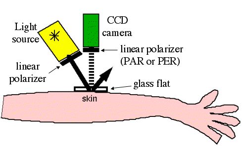
Fig. 1: Simple polarized ligth camera.
Previous page | Next page | Table of Contents
Consider a simple polarized light camera. A white light source passes through a linear polarizer oriented parallel to the source-tissue-camera plane (the scattering plane), illuminating a tissue site. The tissue contacts a glass plate optically coupled with a clear viscous gel. The light is delivered obliquely so that glare from the glass and tissue surfaces reflects away from the camera. The photons scattered from the tissue are collected by a CCD camera after passing through a second linear polarizer, which can be oriented either parallel (PAR) or perpendicular (PER) to the polarization orientation of the illumination light.
The orientation of polarization of the multiply scattered photons from the deeper tissue layers are randomized and contribute equally to both the PAR and PER images. The difference image, PAR - PER, cancels this common background and creates an image based only on the photons scattered just once or a few times which retain the polarization of the illumination light.
The polarized light image emphasizes the structures of the upper 300 um of a birefringent tissue such as skin.
Fig. 1: Simple polarized ligth camera.